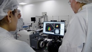
HORIBA Scientific, specialists in measurement and analysis solutions for research and industry, and world leader in Raman spectroscopy, announces combined products using leading technology from CytoViva, Inc. By combining HORIBA’s Raman microscopes with CytoViva’s Hyperspectral Imaging (HSI) microscopy module and Enhanced Darkfield (EDF) illumination, Raman analysis is faster and more powerful.
This innovative integration is of major interest for applications related to nanomaterials research, drug delivery, nanotoxicology studies and SERS nanoparticles characterization. Hyperspectral imaging microscopy allows rapid imaging across the sample with high sensitivity. Colored images generated from the spectra guide the user to easily locate nanoparticles and features of interest.
The patented CytoViva enhanced darkfield illumination improves the signal-to-noise ratio up to ten times over standard darkfield microscopes. The detection limit in size improves sufficiently to allow visualizing nanoparticles as small as 10 nm when isolated.
Raman spectroscopy is a powerful technology for research and industry and provides detailed information on sample properties such as chemical structure, phase, polymorphism, intrinsic material stress, contamination and impurities.
Integrating Raman with hyperspectral imaging and enhanced darkfield allows users to rapidly visualize the sample and target regions of interest. They can then leverage Raman measurements from the identical field of view to provide and confirm the chemical identify of nanoparticles or other sample elements.
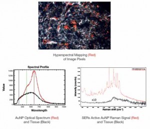
n this application example, gold nanoparticles are imaged in tissue (top), with the corresponding optical (bottom left) and Raman (bottom right) spectra showing clear differences between the nanoparticles and tissue. Visualization of the nanoparticles is made simple with the hyperspectral imaging, while Raman provides a detailed chemical fingerprint.














 Back to Products