Microscopy has been around since Galileo developed a compound microscope in the 1600s. Fast forward to the 1980s when the Nobel Prize in Physics was awarded for work on the electron microscope and for the scanning tunneling microscope. In the 1990s, super-resolution microscopy was developed. In 2014, the Nobel Prize in Chemistry was awarded for the development of super-resolved fluorescence microscopy. In 2017 the Nobel Prize in Chemistry was awarded to the team that developed cryo-electron microscopy. And the advances continue as researchers combine technologies, use microscopy techniques with imaging tools, or develop novel techniques for long-term observation of slow processes.
Combining TIRF and iSIM for fast capture
Researchers have merged two types of microscopy to create sharper and faster images of processes inside of living cells. The two technologies are traditional total internal reflection fluorescence (TIRF) and instant structured illumination microscopy (iSIM). The two methods each have their own drawbacks, but the combination of the two seems to compensate for them.
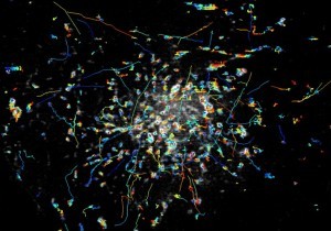
TIRF, which has been around for more than 30 y ears, produces blurry images of small features within a cell. When super-resolution techniques were applied in an attempt to improve resolution, speed had to be sacrificed, so clear images of fast moving objects was out of the question. iSIM was developed in 2013, and it can capture video at 100 frames per second, but it lacks that high contrast that is a feature of TIRF. The combination of the two improves the spatial resolution of TIRF without compromising speed. The result is that the researchers are able to observe rapidly moving objects about 10 times faster than other microscopes at similar resolution (see image above).
For more on the development of the TIRF-iSIM microscope at the U.S. Department of Health & Human Services’ National Institute of Biomedical Imaging and Bioengineering (NIBIB), read TIRF-iSIM Microscope Sharply Images Biological Processes in Living Cells.
Cryo-Electron microscopy reveals workings of herpes virus
Cryo-electron microscopy can deduce the structure of biomolecules by firing beams of electrons at proteins that are frozen. In the UK, researchers have used cryo-electron microscopy to reveal the biological mechanisms the virus uses to infect people.
Cryo-electron microscopy reveals common herpes virus structure
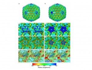
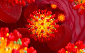
The herpes family uses a capsid or shell to store its DNA and infect its host. And the viruses pump their DNA into preassembled capsids through a motor-like assembly called a portal. That capsid is only 1/10000th of a millimeter in diameter, making it very difficult for scientists to analyze. But using cryo-electron microscopy coupled with new computational image processing methods, the researchers were able to reveal the structure of the portal. They’re hoping that by understanding how the virus p acks its DNA inside the capsule, that it will help to design drugs that will block the action of the portal motor.
For more detail on this study conducted at the University of Glasgow in Scotland, read Cryo-Electron Microscopy Reveals Common Herpes Virus Structure.
Understanding kidney disease with novel force imaging technique
To investigate the function of the kidneys, doctors need to be able to assess the mechanical pressure that the glomerular filter is under. The way the kidneys work is they filter waste products from the blood and remove them in the form of urine, via glomerular blood filtration and a group of cells called podocytes. Mechanical force is key to the survival of podocytes, however, in the past it has been difficult to measure mechanical forces.
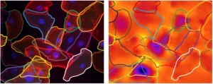
A team at the University of St. Andrews in Scotland used a novel force imaging technique, elastic resonator interference stress microscopy (ERISM), which uses an interference-based approach that requires low light intensity. It facilitates very precise imaging of cellular forces. By using this technique, the researchers found that injured podocytes experience near-complete loss of cellular force transmission. The loss of force is reversible, and so, by using this imaging technique, the patients have a window of opportunity for recovery.
For more on this new technique read Force Imaging Technique Revolutionizes Understanding of Kidney Disease
Written by Anne Fischer, Managing Editor, Novus Light Technologies Today















 Back to Features