As cases of chronic kidney disease continue to rise, scientists now have a better understanding of its causes and treatment, thanks to research led by the University of St Andrews in Scotland (UK).
Glomerular filter under hydrostatic mechanical pressure
The new study, published in Science Advances, has discovered that the glomerular filter, a key component of the functionality of the kidneys, is under substantial hydrostatic mechanical pressure. Kidneys filter waste products from our blood and remove them in the form of urine, via glomerular blood filtration and a group of specialist cells called podocytes. Chronic kidney disease presents with damaged podocytes and a sustained reduction in glomerular filtration rate and is an increasing health burden with limited treatment options.
Mechanical force is crucial to the survival of podocytes. However, podocyte mechanobiology remains poorly understood, largely due to the technical challenges in measuring these forces. Now, an interdisciplinary team from St Andrews has investigated the mechanical forces that podocytes apply when they interact with their underlying substrate.
Elastic resonator interference stress microscopy (ERISM)
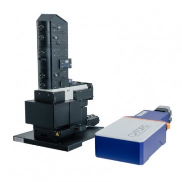
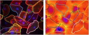
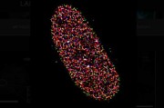
Dr Paul Reynolds from the School of Medicine, in collaboration with physicist Professor Malte Gather, used a novel force imaging technique, elastic resonator interference stress microscopy (ERISM), developed by Professor Gather. Their findings show that injured podocytes experience near-complete loss of cellular force transmission. This loss of force is reversible and as such there is a window of opportunity for recovery from injury.
Investigating the mechanobiology of podocytes
Dr Reynolds says: “Mechanobiology is an emerging area of research and very relevant to kidney disease. We show that contractile podocyte forces are transmitted via specific contact points made by cells on their substrate. We also show the significance of the loss of these mechanical forces in podocyte injury.
“Our work paves the way to a new level of understanding of the kidney by describing the mechanobiology of podocytes. These findings will open new avenues of exploration in the treatment of podocyte injury. Our approach also has general applicability to a wide range of biomedical questions involving mechanical forces.”
Both Dr Paul Reynolds and Professor Malte Gather are part of the University’s interdisciplinary Biomedical Sciences Research Complex.











 Back to News