


Although fluorescence microscopy has played an important role in the recent explosive advancements in live cell imaging (LCI), it still presents challenges. Photobleaching and phototoxicity limit repeat measurements. Rendering high-contrast molecular information requires labelling agents that may interfere with normal molecular activities. Vitally, cells that need to be analysed and then reinserted into the body for in vivo studies, such as stem or immune cells, pose an especially difficult challenge.
Recently, a new 3D imaging technology emerged that provides a quantitative, label-free means to image live cells and tissues. Tomocube’s holotomography (HT), also known as Quantitative Phase Imaging (QPI), delivers nanoscale, real-time, dynamic images quickly and simply without any sample preparation. Now, Tomocube’s latest microscope combines QPI with fluorescence for superior spatiotemporal resolution as well as high molecular specificity HT, revealing the structure, volume, surface area, concentration, and dry matter mass of individual live cells in real-time. This achievement was recognised by the prestigious 2019 Microscopy Today Innovation Award.
How holotomography works
Refractive Index (RI) is a fundamental optical parameter describing the speed of light passing through any object, such as a cell. As the light passes through, individual cellular constituents scatter the light and change its phase according to their individual RI. Phase contrast microscopy picks up these tiny brightness changes as changes in contrast to reveal cellular details.
The Tomocube HT microscope records multiple 2D RI measurements and stitches them together to display a 3D image, or tomogram (Fig. 1--above). Figure 1 shows Hepatocyte detail captured by Tomocube’s HT-1 holographic microscope.
Held between the objective and condenser lenses, the sample is illuminated with a 532nm laser and onto a digital micro-mirror device (DMD) containing several hundred thousand micromirrors. This electronic control of the light-path eliminates moving parts for enhanced stability and image resolution and allows the beam to be rotated around the specimen to collect all the 2D data to be stitched in less than a second (Fig.2).

Schematic light path - Sample is located on the stage between the objective and a condenser lens. A laser is split into a specimen and reference arm. The sample and the reference arms generate a 2-D hologram, which is recorded by a digital camera. Under the control of the digital micromirror device (DMD), the laser illuminates the sample with an incident angle of 53° or 63° rotating 360° with respect to the optical axis. A 3-D refractive index (RI) tomogram of the sample is then reconstructed from the measured multiple holograms with various illumination angles.
Since RI values are quantitative, HT microscopy provides valuable quantitative data about the sample, including morphological and chemical information.
An advantage for live cell imaging
HT microscopy exploits the intrinsic optical properties of all materials, measuring the optical phase delay introduced by RI differences between samples and their medium directly. Its QPI technology only requires a low power laser system, which eliminates the potential for photobleaching and phototoxicity. It also eliminates the need for stains or other invasive labelling agents to investigate live biological material simply and rapidly. The reconstructed 3D tomograms quantitatively resolve structural and chemical information at high resolution, including dry mass, cell volume, shapes of sub-cellular organelles, cytoplasmic density, surface area, and deformability. Its capability to capture 3D images of unlabelled live cells is demonstrated in Fig. 3.

Cellular images captured by the HT-1 holographic microscope.
Main features
- 3-D Refractive Index – quantitative phase imaging of biomaterials
- No labelling or sample preparation – rapid and convenient
- Very low laser power – zero stress on the sample
- High resolution - optical resolution of 110 nm
- Fast - in 2D and 3D
Holotomographic quantitative phase imaging and fluorescence
Although the label-free and high-speed 3D imaging attributes of QPI are highly advantageous for live cell imaging, the limited molecular specificity restricts broader applications in cell biology and biochemistry. This would be overcome if 3D QPI was combined with fluorescence.
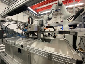
Tomocube has taken this approach with the launch of its second microscope, the HT-2, which adds 3D fluorescence imaging capability to the existing holotomography set-up (Fig. 4).

The Tomocube HT-2 holotomography and fluorescence microscope
The HT-2 is the first instrument of its type and delivers holotomography/fluorescence correlative analysis in 2D, 3D and 4D and enables researchers and clinicians to open new frontiers in bioscience and better understand, diagnose, and treat disease.. The highly detailed fluorescence images show the position of specific target organelles or structures in living cells, while simultaneous measurements of time-lapse 3-D RI tomography enables the long-term tracking of specific targets in live cells while minimising stress.
The HT-2 incorporates a customisable three-channel LED light source (385, 470 and 570 nm) and a motorized Z-drive with a step resolution of 150 nm to generate highly detailed Z-stack images. The TomoStudio software suite controls all the functionality of the HT-2 and provides fast imaging capability and 2D/3D/4D visualization of the cellular images based on 3D RI distributions of the cells and tissues. At its core is the same patented complex digital micromirror device (DMD) optical light shaper used in the Tomocube HT-1.
Benefits
- Correlative microscopy in one instrument - HT-2 provides high-quality 3D images of both holotomography and 3D fluorescence for each sample.
- Quantitative data marked with fluorescence - HT-2 provides morphological (volume, surface area, projection area, sphericity and ellipticity), chemical (dry mass, concentration) and mechanical (cell deformability) properties of cells with 3D refractive index (RI) tomogram. Moreover, fluorescence image provides information about molecular specificity.
- Live cell molecular and holographic imaging with minimal stress on cells - Simultaneous measurement capability of time-lapse 3-D RI tomography and fluorescence image allows long-time tracking of specific targets in live cells. The fluorescence image provides the position of specific target organelles or structures in live cell, and consecutive measurements of time-lapse 3-D RI tomography enables the monitoring of cells and their structures with minimal stress.

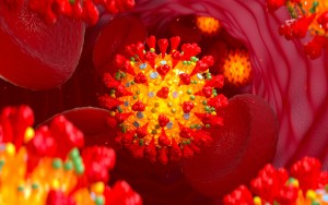
Figure 5A shows time-lapse 3D images of Toxoplasma gondii infecting an ARPE-19 cell, which undergoes death as it is penetrated. The high-resolution time-lapse 3D images can provide important insights on the mechanism of infection and were measured using a Tomocube HT-1 holotomography microscope (HT-1H, Tomocube Inc., Republic of Korea). Fig. 5B shows NIH-3T3 cell research using QPI and 3D fluorescence (GFP-Mito and mCherry-Golgi). Both images were obtained using a Tomocube HT-2 holotomography microscope (HT-2H, Tomocube, Inc., South Korea).
The power of QPI combined with fluorescence in a single instrument is shown in Fig. 5. The high-resolution time-lapse 3D images of Toxoplasma gondii infecting an ARPE-19 cell (Fig. 5A) provide important insights on the mechanism of infection and were achieved using the Tomocube HT-1 microscope. Meanwhile, Fig. 5B shows NIH-3T3 cell research using both QPI and 3D fluorescence (GFP-Mito and mCherry-Golgi) on a Tomocube HT-2 holotomography microscope.
The future’s dual
Holotomography and other QPI techniques have much to offer researchers striving to understand the complex interactions within living cells. The emergence of correlative imaging shown by the award-winning HT-2 microscope from Tomocube, which combines QPI’s speed and quantitative approach with fluorescence’s molecular specificity of fluorescence, strongly suggests these innovative approaches will enhance our understanding of the physiology and pathology of cells and tissues and make a critical impact on the diagnosis and treatment of various diseases.
Written by by Aubrey Lambert, Chief Marketing Officer, Tomocube Inc.