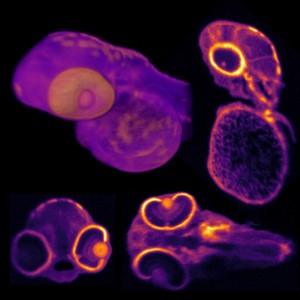
Optical coherence tomography (OCT) uses light for cross sectional imaging and is most commonly used for medical diagnostics, but it also has uses in applications such as analyzing paint on automobiles, nondestructive testing, and more. The global optical coherence tomography market is projected to expand at a CAGR of 15.41% from 2022 to 2030 driven mostly by demand for its use in diagnosing eye disease and other disorders. The COVID-19 pandemic also led to a surge in use as OCT angiography is used for the imaging of retinal vascular changes in fully recovered COVID-19 patients.
OCT has advanced in recent years; however, a few challenges remain. It is difficult to acquire high-resolution OCT images over a wide field of view in all directions, simultaneously. Also OCT images can contain a lot speckle, caused by random noise.
3D optical coherence refraction tomography
In attempt to resolve these challenges, researchers at Duke University developed a new OCT technique called 3D optical coherence refraction tomography (3D OCRT), which produces highly detailed images that would be difficult to observe with traditional OCT.
“We developed a new and exciting extension, featuring novel hardware combined with a new computational 3D image reconstruction algorithm to address some well-known limitations of the imaging technique,” said Kevin C. Zhou, lead author of the study.
Parabolic mirror
What was unique to their setup was that they used a parabolic mirror, which made it possible to image a sample from multiple views and a range of angles. They developed an algorithm that combines the views into a single, high-quality, 3D image that is corrected for noise and other distortions.
“We envision this approach being applied in a wide variety of biomedical imaging applications, such as in vivo diagnostic imaging of the human eye or skin,” said research team co-leader Joseph A. Izatt. “The hardware we designed to perform the technique can also be readily miniaturized into small probes or endoscope to access the gastrointestinal tract and other parts of the body.”
The researchers were able to overcome engineering challenges they had faced in previous studies. "Because our system generates tens to hundreds of gigabytes of data, we had to develop a new algorithm based on modern computational tools that have recently matured within the machine learning community,” said Sina Farsiu, research team co-leader.
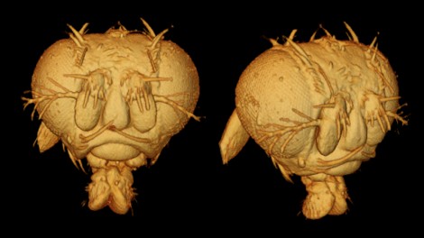
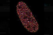
The researchers imaged biological samples including a zebrafish, fruit flies (shown here), mice, and more, and were able to acquire 3D fields of view of up to 75 degrees without moving the sample.
“In addition to reducing noise artifacts and correcting for sample-induced distortions, OCRT is inherently capable of computationally creating contrast from tissue properties that are less visible in tradition OCT,” said Zhou. “For example, we show that it is sensitive to oriented structures as fiber-like tissue”
Paper: K. C. Zhou, R. P. McNabb, R. Qian, S. Degan, A.-H. Dhalla, S. Farsiu, J. A. Izatt, “Computational 3D microscopy with optical coherence refraction tomography,” 9, 6, 593-601 (2022).
DOI: 10.1364/OPTICA.454860











 Back to Features

