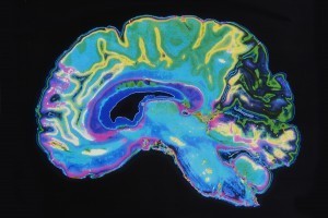
Introducing three new optical windows in the near-infrared (NIR) region, researchers at The City College of New York (CCNY) in the US have for the first time created a “Golden Window” for deep brain imaging and proved that using light at wavelengths 1600–1870 nm enables more noninvasive, more detailed study of the brain.
What is the “Golden Window” for deep brain imaging?
“One of the major thrusts in neuroscience is to image deep inside the brain with micrometer or sub-micrometer resolution,” explains Dr Lingyan Shi, who is leading deep brain imaging research in CCNY’s Institute for Ultrafast Spectroscopy and Lasers. Optical imaging is still the only method to study neural tissues with such high resolution. Compared with visible light, light in the NIR spectral zone has the advantage of reduced absorption and scattering, which promotes a less blurred image. The expert says the experimental and theoretical results show that the Golden Window — covering wavelengths 1600–1870 nanometers — is an optimal window for light penetration in brain tissue, followed by Windows II (1100–1350 nm), IV (centered at 2200 nm) and I (650–950 nm). “It is evident that brain tissues become more transparent when using the light of the Golden Window,” she adds.
NIR optical windows and mechanisms of deep tissue penetration
According to Shi, researchers have experimentally reported that light in Window III potentially penetrates deeper in tissue, which is consistent with the present study of the brain. “But they did not look into the mechanism or compare with Window IV, which is easily presumed to be superior to Window III,” the biomedical engineer says, adding that her team’s study is the first to explain why Golden Window light can go deeper in brain and what the mechanisms of deep penetration in tissue are.
In her institute, noninvasive imaging of the brain is mainly for laboratory study on small animals, such as rodents, whose skulls are relatively thin. However, “It is also possible to obtain deep imaging in breast or prostate tissue or other types of tissue,” Shi says. “When both the excitation (laser wavelength) and emission (dye) wavelengths fall within the Golden Window, imaging depth could be further optimized.”
The expert agrees that her team’s breakthrough in light technology could have noteworthy impact on the development of the next generation of microscopy imaging techniques. “Laser scanning microscopes are major instruments used by researchers in the neuroscience community,” she says. “If the new laser can provide wavelengths in the Golden Window, deeper layers of brain cortex imaging with sub-micrometer resolution may be achieved with single-photon excitation or two- or three-photon excitation... or Second Harmonic Generation (SHG)/Third Harmonic Generation (THG),” explains Shi.
As for herself and her colleagues, she says the next step is to advance time-resolved imaging to primary events in brain activity.
The study “Transmission in near-infrared optical windows for deep brain imaging" is published in the Journal of Biophotonics.
Written by Sandra Henderson, research editor Novus Light Technologies Today











 Back to Features

