


Phase Focus Ltd (Phasefocus) announces a family of phase microscopy systems for live-cell imaging using the Phasefocus Virtual Lens (VL). The VL acquisition and processing engine uses a technique known as "ptychography" to provide quantitative, high contrast, label-free images of cells for long-term time-lapse studies.
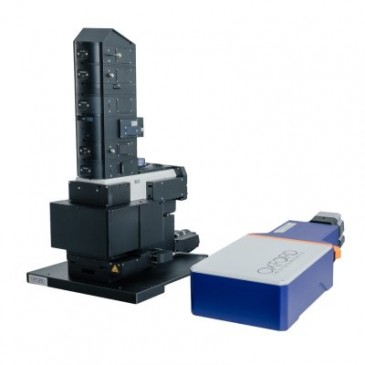
The use of fluorescent probes is commonplace for labelling fixed specimens. However, when used in living cells, fluorescent probes can be toxic and ultimately disturb the natural cell function. The use of laser illumination can also be phototoxic to cells. The Phasefocus VL20 upright and VL21 inverted live-cell imaging systems use low-power laser illumination, enabling long-term time-lapse cell imaging including multi-area time lapse to produce high-contrast quantitative image information.
In addition, the quantitative nature of the phase information means that data such as cell thickness, cell volume and cell motility can be measured on a familiar and easy-to-use system for the study of both individual cells and cell populations.
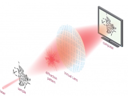
Schematic of the Virtual Lens
The VL is based on technology developed at the University of Sheffield by Professor John Rodenburg. It provides an iterative solution of the diffraction pattern phase problem and is applicable to visible light imaging, x-ray imaging and atomic resolution electron-imaging. Phasefocus has applied the technology to develop and install two systems at the University of York in the Department of Biology as one of many technology-leading techniques in the Imaging and Cytometry group managed by Dr. Peter O’Toole.
Dr. O’Toole describes ptychography as "one of the most important breakthroughs in imaging." (For more specifics on ptychography, see the article "Phase Focus Issues Paper on Ptychography" on Novus Light Technologies Today.)