Researchers at Tel Aviv University, Israel, have combined commercially available hardware and the open-source software PySight to improve the quality of rapid 2D and 3D imaging of neuronal activity in the living brain.
How PySight was born
The laboratory of Pablo Blinder, PhD, at Tel Aviv University, strives to uncover the inner workings of the interface between neurons and blood vessels. Oxygen and nutrients are distributed to active areas on a need-basis, which is not only important for proper brain function but is also the underlying mechanism of functional magnetic resonance imaging (fMRI), which cannot “see” neuronal activity but rather its signature on blood flow.
According to Blinder, both systems need to be recorded simultaneously to decipher the transforming function between the neuronal activity and the vascular output. 2D imaging misses what his team believes to be crucial information necessary to understand this process at the cellular and sub-cellular levels. “So we went into a quest to volumetrically image these dynamics across the depth of a mouse cortex — there is a depth or layer-dependent behavior that is part of the whole story,” he notes. It became quite clear to the team that existing tools were not up to the task: “We wanted to use the fastest continuous volumetric imaging available and further extend the size of the volumes of interest. When you try to do so, you enter into the photon-deprived regime. Under this condition, only photon counting — as opposed to analog integration — can do the trick. At that moment, our quest was set: to build a volumetric imaging solution based on photon counting.”
Creating an easy-to-integrate photon counting solution
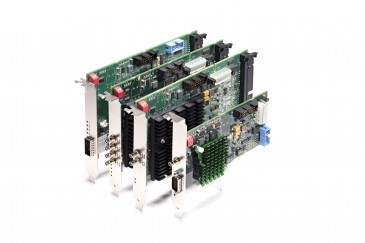
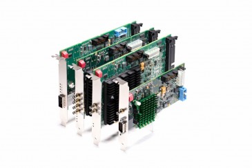
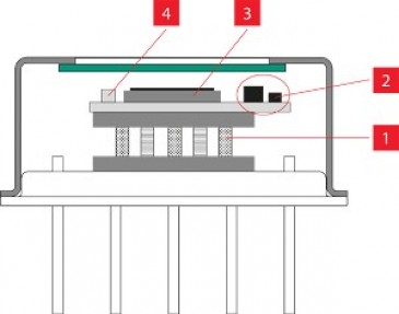
Photon counting is a decades-old method, but Blinder points out that DIY setups require some heavy lifting in terms of electronics, limited to “excellent laboratories” with the adequate know-how. Commercially available systems did not even come close to what Blinder and his colleagues needed. “It became clear that developing an easy-to-integrate photon counting solution could benefit many laboratories in the multiphoton microscopy community, and this is exactly what we set out to do,” the research leader says. “With the help of Professor Ori Cheshnovsky, we designed the concept of PySight, which is based on time-correlated single-photon detection. In addition to storing the photon-detection times, we also store timing signals from the scanning elements.” They used a commercially available multiple-event time digitizer — or multiscaler — and built PySight as open-source software around it to create an add-on that can easily be integrated into any multiphoton microscope as long as the PMT and scanning timing signals can be “hooked” into the multiscaler, according to Blinder. PySight can also be used to parse data from other systems using its API.
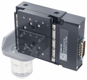
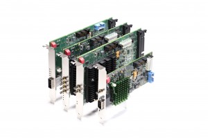
“Another important issue is that PySight allows us to do unsynchronized volumetric scanning using a varifocal lens, or TAG lens,” Blinder adds, explaining that doing so eliminates the need for synchronization electronics. “Putting everything together, PySight, hardware selection, which is top performing, and its software are a true plug-and-play solution for any microscopist seeking to image faster and over increasingly larger tissue volumes.”
Improved imaging under photon-deprived conditions
“It turns out from our measurements that the most commonly used approach to imaging neuronal activity with the help of genetically encoded calcium indicator — even in 2D and at 30 frames per second — already set every lab performing this technique into the photon-deprived conditions,” Blinder emphasizes. “We showed that the use of photon counting through PySight significantly improves the detection of neuronal activity.” Moreover, more accurate genetically encoded voltage membrane indicators required trail averaging under analog acquisition, and the Tel Aviv team showed they could detect the signals in a single trail. “We attribute, potentially, this improvement to the removal of a specific noise that contaminated the output of the detector,” Blinder says, reasoning it had been well known photon counting can remove this noise.
Portability and easy integration
Aiming to demonstrate portability and easy integration, Blinder reports his team walked across campus to the lab of Dr Moshe Parnas, who studies odor coding in the fly brain using a commercial multiphoton microscope. “It was a matter of a few minutes to connect [and] rewire signals, download software, and we got it working to record signals from a genetically encoded voltage indicator.”
Ultrafast continuous volumetric imaging
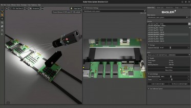
PySight allows easy integrating of the fasted axial scanning solution without the need for additional electronics to sync with the existing scanning system. “We now used it to record cortical neuronal activity in an awake mouse in a volume of 685 x 880 x 270 μm³ at 30 volumes per second,” Blinder says. (Watch the video).
The research is described in the paper “PySight: plug and play photon counting for fast continuous volumetric intravital microscopy,” published in Optica.
Written by Sandra Henderson, Research Editor, Novus Light Technologies Today