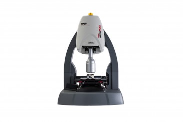


Long-term studies of live cells are set to take a significant leap forward with the introduction of a motorized stage and custom-designed stitching software for Tomocube’s holotomography microscopes. The automated combination can be fitted to every microscope model in the company’s range and produces large field-of-view images and time-lapse videos.
The company says the combination of holotomography microscopy and the new motorized stage and stitching software is potentially a game-changer for the field of live cell imaging. Only a low-power laser is needed to image live cells and tissues in three dimensions without stains or fluorescent probes. So researchers can now conduct long-term studies without the fear of photobleaching and phototoxicity impacting their results.
According to Tomocube, even when the studies are conducted on the new Tomocube HT-2, which combines holotomography and 3D fluorescence imaging into one unit for the first time, the potentially damaging fluorescence observations can be restricted to mapping molecular specificity while relying on the gentler holotomographic quantitative phase imaging for the longer-term observations of the cells.
Tomocube says they are the first company to fit a motorized stage to a holotomography microscope and have worked with a team from the Korea Advanced Institute of Science and Technology (KAIST) to develop the custom stitching software for the motorized stage. With a step size of 70 um, the stage allows specimens up to 8 mm x 8 mm to be imaged in their entirety. The motorized stage is equipped with “Mark & Find,” which enables multiple cell locations to be recorded and rapidly retuned to throughout an experiment, and “Click & Shift,” which centers a defined target position in the field of view. Fast image acquisition of holotomographic images on the HT-1 and HT-2 microscopes allows 2D and 3D images to be captured at rates of 150 and 2.5 frames per second respectively across the entire 8 mm x 8 mm stage acquisition area.
This integration means that all the possibilities offered by a large field-of-view in live cell and tissue research are now available for the first time without any sample preparation, staining or labelling. What is more, it provides structural and chemical information from the cells at high resolution, including dry mass, cell volume, shapes of sub-cellular organelles, cytoplasmic density, surface area and deformability.
Specifications
- Step size : 70 um x 70 um
- Maximum FOV (field of view) of single acquisition: 80 um
- Motorized stage acquisition area: 8 mm x 8 mm
- 2D acquisition: 150 fps
- 3D acquisition: 2.5 fps.