Light technologies are increasingly contributing to our health and longevity. While, for example, cancer is among the leading causes of death worldwide, death rates for individual cancer types have declined in part due to advanced research on light technologies such as fluorescent imaging that helps in cancer detection and photodynamic therapy used in treatment. In other research, lighting is found to increase alertness and help with sleep, which increases overall health. Optical coherence tomography devices are tweaked for arthroscopic use. Researchers in Austria are using two-photon polymerizataion method to develop a high-resolution bioprinting process capable of 3D printing living cells. And mass spectroscopy is used to detect photoinitiators in breast milk, and to assess the rate of consumption and potential risk for infants.
NIR-II fluorescence endoscopy for targeting colon cancer
A multidisciplinary research team in China and the US collaborated on the design and construction of an endoscopic system that can perform real-time non-invasive imaging of colorectal tumor biomarkers. What’s important about this work is that they are the first to report the use of NIR-II fluorescence endoscopy for the targeted detection of colorectal cancer. The system they developed enables the simultaneous acquisition and display of white light and NIR-II fluorescence, and it delivers subcellular resolution of 20 μm for sharp images in the NIR-II region. Read more at NIR-II Fluorescence Imaging of Colorectal Cancer.
The effect of light on alertness and sleep
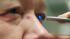
At the Lighting Research Center at Rensselaer Polytechnic Institute in Troy, New York (US), researchers studied how specific light promotes circadian entrainment and alertness in the office environment. What’s significant in this work is that it is the first to demonstrate that red light in combination with ambient white light helps make people more alert. Specially designed luminaires were mounted near the participants’ computer monitors and objective and subjective measures were used to evaluate the effects of the lighting. It was found that morning blue light advances the circadian phase, enabling participants to wake up earlier in the morning. Afternoon red light made participants more alert, reducing sleepiness and increasing vitality and energy. Read more at Morning Blue and Afternoon Red Light Increases Alertness in Workers.
Organic molecules and quantum confined silicon nanocrystals work together to fight cancer
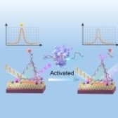
Materials scientists in Texas and California (US) have pioneered how to attach conjugated organic molecules to silicon nanoparticles. What is significant about this is that while it has been known that high energy light can form free radicals that can attack cancer tissues, scientists have wanted to use silicon because it is non-toxic. But until now, silicon nanocrystals have not been able to up-convert photons—the process that is necessary to get free radicals to attack cancer tissue.
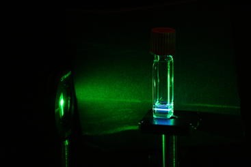
This group learned how to attach ligands to the nanoparticle that are specifically designed to transfer the energy from the nanocrystals to surrounding molecules. They used triplet fusion to turn low-energy photons into high-energy photons, ultimately pioneering how to attach conjugated organic molecules to the silicon nanoparticles. Read more at One Step Closer to Minimally Invasive Photodynamic Treatments for Cancer.
Compact OCT device in arthroscopic surgery
Optical coherence tomography (OCT) is a standard in ophthalmology, however, making an OCT instruments compact enough for imaging inside of other areas of the body has been a challenge—until now. Researchers from Duke University wanted to develop an OCT device that could be used in arthroscopic surgery. To make such an OCT system, the researchers created an endoscopic delivery system that uses a rigid 4mm-wide borescope to relay the image from the tissue to a fiber optic connection.
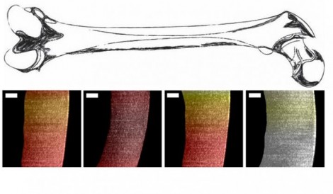
The result is an OCT instrument that provides real-time information on cartilage thickness without requiring the surgeon to cut any tissue. The system was able to identify the bone-cartilage interface for samples that were less than 1.1 millimeters thick. The next step is to demonstrate that it can image thicker samples, as well as improving the ergonomics for use in surgery. Read more at Low-Cost Portable System Takes OCT Beyond Ophthalmology.
Photoinitiators used in food packaging found in breast milk
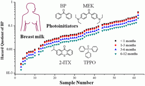
Photopolymerization is considered a “green” technology used in the manufacturing of light-sensitive materials, such as UV-curable inks that are applied in packaging materials. Generally, the photoinitiators (PIs) that are used break down; however, scientists have detected them in food, indoor dust and blood serum. Recently, researchers at the University of Toronto used mass spectroscopy to detect PIs in breast milk.
Living cells produced by 3D printing method
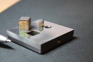
It seems that materials and objects of all types can be printed by a 3D printer, including living cells. However, it is not without its challenges, as there has been a lack of suitable chemical substances. A team at TU Wien in Vienna (Austria) used an extremely high-resolution, two-photon polymerization method to develop a high-resolution bioprinting process with complete new materials.
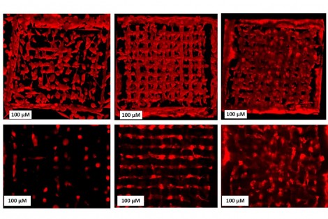
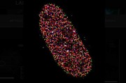
They have developed a “bio ink”, which enables cells to be embedded in a 3D matrix printed with micrometer precision at a speed of one meter per second. The polymerization method requires a high-intensity laser beam, which can also be used to determine how easily the structure will degrade over time. The technique of “bioprinting” embeds the living cells directly into the 3D structure during the production of the structure. The image above shows cells spreading in a 3D scaffold. Professor Aleksandr Ovsianikov, head of the 3D Printing and Biofabrication research group at the Institute of Materials Science and Technology said, "Using these 3D scaffolds, it is possible to investigate the behavior of cells with previously unattainable accuracy. It is possible to study the spread of diseases, and if stem cells are used, it is even possible to produce tailor-made tissue in this way". Read more at Bioprinting Living Cells in a 3D Printer.
Written by Anne Fischer, Managing Editor, Novus Light Technologies Today









 Back to Features