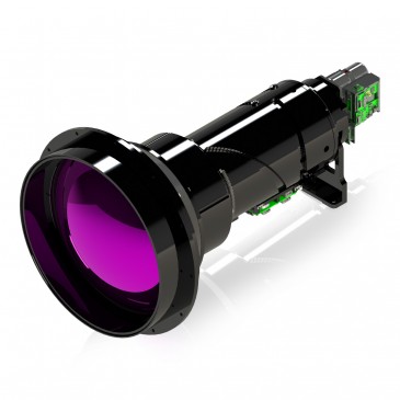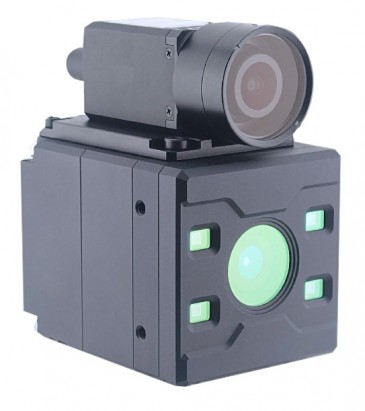


A custom portable optical imaging system developed at the University of Illinois at Urbana-Champaign (UIUC) visualizes the dynamic cellular and molecular features in freshly removed or biopsied breast tissue specimens, as opposed the typical several days current tissue pathology and diagnostics take.
Images of molecules and cells in tissues without dyes or labels
“Our optical imaging system generates images of molecules and cells in tissues based on multiple nonlinear optical processes, without the use of any dyes or labels that would perturb the dynamics,” says UIUC bioengineering professor Stephen Boppart. The new optical imaging system is portable and cart-based, which enabled the researchers to use it in the operating room to assess the described dynamics in freshly resected breast tissue and tumor specimens in real time. The team was also able to image and quantify extracellular vesicles from their images, which the professor says have direct implications in carcinogenesis and cancer staging as a biomarker.
A new kind of imaging technique
“In our system we use a particular excitation wavelength that allows four nonlinear excitation processes to occur, without having the emission signals overlap,” Boppart says. “This provides both structural and molecular imaging, all without the use of labels.”
The researcher says they made their system portable for intraoperative use of fresh tissues. “And with it,” he continues, “we could uniquely image and quantify the extracellular vesicles.” Boppart adds that extracellular vesicles, including exosomes, are an active area of research investigations. “We believe this is the first demonstration of detecting or quantifying these in fresh tissues in the operating room.”
Histology-level images in real-time in the OR
The new optical imaging system solves multiple problems: “First, our label-free strategies eliminates the need for dye or stains that take time to use and can perturb the dynamics and functions of the cells in tissue,” the professor says. “Second, we can generate histology-level images in real-time in the OR instead of waiting hours to days for the standard stained histopathology. Third, we can detect and quantify the extracellular vesicles, which can be used as a biomarker and cancer staging in the OR in real-time.”
The advantages of the portable optical imaging system compared with previous platforms and methods include label-free imaging, multimodal imaging, fast real-time imaging of fresh tissues, and that it can detect and quantify extracellular vesicles.
Other applications
Aside from real-time cancer diagnostics, Boppart says there are potential other applications for this new type of portable optical system. “This methodology can potentially augment or replace histopathogy or immunohistochemistry by providing histology-level imaging at the point-of-procedure and in real-time,” the bioengineering expert says. “This can also be performed in-vivo, potentially replacing the need to take out tissue specimens for biopsy.”
The research breakthrough could have an impact on the development of future optical technologies: “We believe this methodology and technology will more rapidly lead to intravital label-free nonlinear imaging to visualize and understand a wealth of new information on molecular and cellular dynamics,” Boppart says. “Developing new supercontinuum light sources with pulse shaping (coherent control) will also expand our ability to generate new forms of contrast from cells and tissues.”
Next steps
Boppart says he and his colleagues are building new beam-delivery systems to work with this platform, including new fiber-based catheters and endoscopes and hand-held imaging probes. They are also planning to demonstrate they new optical imaging system at the point-of-procedure for assessing dynamics in needle-biopsy specimens. Furthermore, Boppart reveals he has spun-out this technology to a start-up company — LiveBx — which stands for “Living Biopsy.” “We believe there is much to gain from imaging living biopsy specimens before they are killed for standard histopathology processing.”
Written by Sandra Henderson, Research Editor, Novus Light Technologies Today