Leica Microsystems, a leader in microscopy and scientific instrumentation and advanced imaging solutions, announced today that it has acquired ATTO-TEC, a leading specialty supplier of fluorescent dyes and reagents. The addition of dyes and reagents for sample preparation complements the renowned Leica portfolio of microscopy imaging platforms and advanced AI-based analysis software.
“With the acquisition of ATTO-TEC, Leica Microsystems is now able to support researchers at every stage of the microscopy imaging workflow," said Dr. Annette Rinck, President of Leica Microsystems. "This can be a key advantage for reliable results, such as in high-plex 3D experiments in cancer research. Our combined expertise will help researchers to reveal the invisible, enable discovery, and ultimately lead to breakthrough research, accelerating therapy development for improving human health.
Dr. Jörg Reichwein, CEO of ATTO-TEC GmbH, added: "I am convinced that by becoming an integral part of Leica Microsystems, we will mobilize the right forces to differentiate the microscopy imaging offering further. Direct access to knowledge of subsequent imaging and analysis steps leads to new approaches in developing assays, kits and dyes optimised for the entire workflow".
ATTO-TEC will continue to provide its high-quality services and renowned ATTO dyes, antibody labeling kits, labeled phospholipids, and other reagents. ATTO-TEC products will remain available through the ATTO-TEC online store and existing partners.
ATTO-TEC dyes have become a benchmark for fluorescence microscopy imaging, offering a highly differentiated panel. Their brightness and photostability make them the reagents of choice for demanding applications. Notable examples include ATTO 488 and the cornerstone of super-resolution modalities, ATTO 647N.
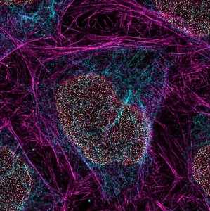
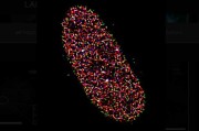
IMAGE: Super-resolution image of cleared zebrafish heart tissue taken with a STELLARIS confocal microscope with TauSTED Xtend. The magenta color shows actin filaments, stained with ATTO 647N dye. Sample Courtesy of Dr. Mariano Gonzales Pisfil and Dr. Steffen Dietzel from the Biomedical Centre at Ludwig-Maximilians-University Munich, Germany.










 Back to News

