


A UK-wide research team, led by the University of St Andrews, has developed an innovative new way to optically image through tissue, which could allow for a more detailed understanding and diagnosis of the early stages of various diseases, including cancer.
The study, in collaboration with the University of Southampton and the Cancer Research UK Edinburgh Centre at the University of Edinburgh, published in Science Advances (Friday 12 October), paves the way to move from superficial to functional imaging, transforming studies in neuroscience.
Light is incredible. Its ability to image objects has had a profound impact across all of the sciences. However, as we know from everyday experience, light does not penetrate through our skin or a piece of Sellotape very well. The light scatters and is scrambled. This in turn makes it very hard to create images from deep within a sample.
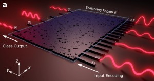
The innovative new method developed by the team of researchers focused short pulses of patterned light in time through the tissue. By focusing in time, known as temporal focusing, the patterns retain their form despite the scattering from the tissue. However, this is not imaging. To image, the team collected just a fraction of the return light (fluorescence) from the sample onto a single-point detector. This means they did not have to know where that light came from within the sample. By simply appropriately summing the patterns projected on the sample weighted by the intensities recorded for the return light, the team were able to form a faithful image. Crucially this image was created without ever having any specific knowledge of the tissue itself.
The ability to see deeper into tissue with light is currently one of the hottest topics in imaging. The potential applications of the research findings could have wide-ranging implications to aid biomedical analysis and early detection of diseases, including furthering our understanding of neuroscience and degenerative brain diseases.
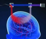
Caption: Laser light patterns are sequentially focused in time onto a region of interest inside biological tissue. Fluorescence emitted by the sample under each illumination pattern is collected with a single-pixel detector after passing back through the tissue. By adding up the projected patterns weighted by the intensities recorded, an image of the sample can be reconstructed
Paper: The paper Wide-field multiphoton imaging through scattering media without correction is published in Science Advances and is available online: http://dx.doi.org/10.1126/