


Although they may be the most bio-diverse marine ecosystems in the world, coral reefs located in tropical oceans near the equator are currently under threat from warming waters, overfishing and water pollution. All these factors compromise the ability of the corals to grow, reproduce and thrive.
Now, a team of researchers at the University of Bristol in the UK are developing a hyperspectral camera that will enable them to develop a greater understanding of these complex ecosystems, and determine how they may be affected by future changes in the climate of the planet.
Inside modern reef building corals (or hermatypic corals) live single-celled algae called simbiodinium -- better known as zooxanthellae -- with which the corals have a symbiotic relationship. The zooxanthellae algae produce oxygen, sugars, amino acids and lipids through the process of photosynthesis that the coral requires to live. In return, the coral produces carbon dioxide, nitrates and phosphates that the algae needs to undertake photosynthesis.
One of the fascinating properties of both the coral and the zooxanthellae is that they both glow -- or fluoresce -- by absorbing one wavelength of light and re-emitting that light at a lower wavelength, producing magnificent colors characteristic of healthy growth. However, the fluorescence of both the coral and the zooxanthellae is downgraded when the coral is stressed as well as injured or damaged.
Turning white
Speaking at the Hyperspectral Imaging and Applications Conference held in Coventry, UK on October 10th this year, Jonathan Teague, a PhD Researcher at the University of Bristol, said that there are several ways that hyperspectral imaging can be used to help determine the health of corals in the marine environment.
First, the spatial distribution of the different coral types and species can be analyzed to provide an indication of the population of the coral reef at any given location. Second, researchers can analyze the rate at which corals are being bleached, an effect which causes corals to turn white when they expel the zooxanthellae living in their tissues as a response to an environmental stressor such as temperature rise. Lastly, the fluorescence of the corals can be measured, a process that may serve as an early indicator of the declining health of the coral population.
Hyperspectral imaging has already been employed effectively in the spectral identification of benthos, the community of organisms that live on, in, or near the seabed. Each of the benthic organisms on or near the seabed has a characteristic optical fingerprint. By capturing hyperspectral images of the benthos, the optical fingerprints of the individual organisms can then be determined through software analysis.
This was an approach taken by Tamir Caras from the Ben-Gurion University of the Negev in Israel who, together with his colleagues, used an above- water push broom Spectral Camera by Specim Systems to capture such images.

Figure 1: Hyperspectral imaging can be used to classify the habitat of corals. (Credit Caras and Karnieli, 2015)
After image capture, software (in the form of a support vector machine) was used to automatically classify both the organisms, such as hard and soft corals, as well as inorganic features such as sediment and rocks and determine their distribution (Figure 1).
Imaging frags
With the chlorophyll fluoresces of the zooxanthellae, light energy absorbed by the chlorophyll molecules can undergo one of three fates. It can be used to drive photosynthesis, with the excess energy dissipated as heat, or re-emitted as light – chlorophyll fluorescence. This type of fluorescence can be measured to determine the number of zooxanthellae algae present within the coral, which is then also an indicator of how bleached the coral is.
To determine whether useable spectral data could indeed be obtained from a set of coral samples, the Bristol Researchers spent time at the London aquarium with aquarist Jack Willans to perform some preliminary testing. In the back of the aquarium is a quarantine section where a number small coral fragments (or “frags”) are grown. Using a Headwall Photonics Nano Hyperspectral camera, the researchers captured images of the corals that were growing in the aquarium. The images were then processed with ENVI (Environment for Visualizing Images) Hyperspectral Image Analysis and Processing Tools, which were developed by GEO University, an Estonian company based in Tallinn.
Having processed the images of a number of coral species, the researchers were able to analyze the intensity of the florescence from the corals at specific wavelengths. They could then determine at what wavelengths the fluorescence was caused by fluorescence of the coral and what fluorescence was caused by the zooxanthellae algae (Figure 2).

Figure 2: University of Bristol researchers analyzed the intensity of the florescence from corals at specific wavelengths. They could then determine at what wavelengths the fluorescence was caused by fluorescence of the coral and what fluorescence was caused by the zooxanthellae algae. (Credit J Teague 2018)
To validate whether the use of a hyperspectral camera could also be used to document the affects of coral bleaching, Mr. Teague and his team then took individual coral fragments from the London aquarium (which represented coral species from many different geographic regions around the world) and placed them into a separate, smaller tank. The temperature of the water in the tank was then increased in 2C increments starting from around 26C to 30C, the temperature at which bleaching would typically occur. The temperature increases were set a one week intervals as rapid temperature rise would have killed the coral.
The different coral types in the tank were excited using a 405nm LED lamp and the images of the coral captured again using the Headwall Photonics Nano Hyperspectral camera. The software analysis of the hyperspectral images was again performed to produce emission spectra for each of the corals over the course of the experiment as the water temperature was elevated.

Figure 3: As temperature increases, both the host fluorescence and the chlorophyll fluorescence decrease. (Credit J Teague 2018)
From the emission spectrum, both the host fluorescence and the chlorophyll fluorescence could then be determined. From the experiment, it was clear that as temperature increases, both types of fluorescence decreases and could clearly be detected by the camera (Figure 3).
Cost reduction
Due to the relatively high cost associated with commercially available hyperspectral cameras and the associated risk of underwater applications, the researchers have now created a more practical lower cost (<£10k) push broom hyperspectral camera that can potentially be used for measuring coral health in the marine environment. At its heart is the Atik Horizon from Atik Cameras, a CMOS monochrome camera with a 16MPixel CMOS sensor that was originally developed for deep sky astrophotography.
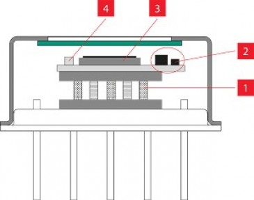
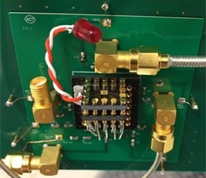
This camera was selected for its large sensor size and relative low cost. To the camera, they fitted a Bifrost LVLWP (Linear Variable Long Wave Pass) filter from Delta Optical Thin Film divided into spectral lines from 450-650nm in 2nm intervals, which can easily convert a ‘normal camera’ into a hyperspectral capable one. This filter sits on a 3D printed mount directly above the chip sensor (Figure 4).

Figure 4: Researchers have now created a low cost (<£10k) push broom hyperspectral camera, which is fully waterproof (<100m) that can be used for measuring coral health in the marine environment and incorporated onto an ROV. (Credit J Teague 2018)
The camera itself will soon be mounted on a BlueROV2 remotely operated vehicle from Blue Robotics to collect data, which can then be used to evaluate the health of coral reefs. As the camera moves over the reef, the push broom scanning camera will enable image data from hundreds of wavelength bands to be recorded at the same time, after which a two-dimensional spectral image of the reefs can be captured and analyzed.
Aside from enabling the Bristol University researchers to measure the distribution of the coral types, build a spectral library of coral fluorescence and bleaching levels, it will also enable them to quantify the concentrations of zooxanthellae present in the corals.
Written by Dave Wilson, Senior Editor, Novus Light Technologies Today