


The use of fluorescence technologies in dental clinical practice is becoming more commonplace as the available range of fluorescence diagnostic devices increases, and the relative cost of these devices reduces. Fluorescence diagnostic technologies provide an important adjunct to conventional dental clinical examinations which relies upon visual examination, probing the surfaces of teeth and into periodontal pockets, and taking dental X-rays. While the conventional methods have very high specificity - being able to exclude the presence of common dental diseases such as dental caries (tooth decay) and periodontitis (gum disease), they have poor sensitivity and are unable to detect many of the earlier forms of destruction which are suitable for less invasive interventions.
More than a dozen manufacturers currently supply the fluorescence diagnostic devices for dentistry. The most widely used types of devices (Table 1) relate to the diagnosis of dental caries, relying on the fluorescence signal generated by bacterial porphyrins to indicate the presence of infection between teeth or within the crowns of teeth. And more recently, similar technology has been developed to allow dentists to assess their work during root canal treatment, a technique that relies upon special fibre optic modifications to provide the correct distribution and collection of light within the confines of the root canal environment.
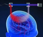
Pulsed laser approach
Because approximately one in five adults suffer from the more aggressive forms of periodontal disease, there is growing interest in the use of fluorescence diagnosis systems to provide an endpoint to the scaling and root planing procedures that are undertaken by dentists and dental hygienists. Such systems can also be coupled to pulsed lasers to allow an autopilot approach to removing bacterial biofilms and bacterial calcified deposits. A similar autopilot guidance strategy can be applied for the removal of infected tooth structure prior to filling a tooth that has undergone root canal treatment.
Switching the view with light
Fluorescence systems also exist that are built into conventional dental equipment such as air turbine drills and scalers, allowing the dentist to easily switch their view of the oral tissues from conventional white light through to visible light induced fluorescence. Several manufacturers provide "lipstick" size miniature intra-oral or cameras that can likewise switch between white light and either blue or ultraviolet light, to enhance discrimination during a dental examination. The same concept has also been applied in the forensic examination of deceased persons in order to establish their identity from dental records, given that many people now have tooth coloured filling some which are difficult to detect under normal white light - it can be easily using fluorescence imaging (Figure 1).
In light of the high morbidity and mortality of oral cancer, considerable effort is being deployed into assessing the usefulness of fluorescence devices for assisting in the clinical diagnosis of both the early and advanced forms of oral cancer. The literature thus far indicates that fluorescence systems can help the dentist in their identifying that had normal oral mucosal is present, but the technology in its current form does not distinguished well between the different types of pathology which may be present (e.g,. an ulcer from trauma, versus an area of cancer). Nevertheless, because fluorescence technologies are safe for both dental clinicians and patients, their use is increasing at a greater rate compared to the various types of dental X-rays.
Table 1. Major fluorescence applications in dentistry: key detection targets
| Dental caries in the fissures of molar teeth |
| Dental caries between the surfaces of molar teeth |
| Decayed dentine within teeth during cavity preparation |
| Dental calculus in subgingival areas in periodontal pockets |
| Infections within root canals of non-vital teeth |
| Tetracycline drugs causing developmental discolouration of teeth |
| Oral cancer and pre-cancer |

Figure 1. Top, white light view of the teeth and oral structures during a dental examination. No unusual features can be seen. Bottom, the same field viewed under UVA 405nm fluorescence excitation with an orange filter. The presence of porcelain crowns (A), deposits of dental plaque and dental calculus on the teeth and near the gingival tissues (B), and a tooth-coloured resin composite filling (C) are readily apparent, because of lack of fluorescence, red fluorescence and bright yellow fluorescence, respectively. Images taken with a custom built multi-wavelength fluorescence dental diagnostic camera (GC Corporation, Japan).
Written by Laurence J. Walsh, Dean of the University of Queensland Dental School (Australia) and a program director within the University of Queensland Centre for Biophotonics and Laser Science.