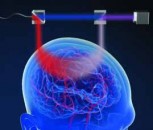


A new optical device puts the power to detect eye disease in the palm of your hand. The tool, which is about the size of a handheld video camera, scans a patient’s entire retina in seconds and could aid primary care physicians in the early detection of a host of retinal diseases including diabetic retinopathy, glaucoma and macular degeneration. Researchers at the Massachusetts Institute of Technology (MIT) describe their new ophthalmic-screening instrument in a paper entitled “Handheld Ultrahigh Speed Swept Source Optical Coherence Tomography Instrument Using a MEMS Scanning Mirror” published in the open-access journal Biomedical Optics Express, which is put out by The Optical Society (OSA).
Although other research groups and companies have created handheld devices using similar technology, the new design is the first to combine cutting-edge technologies such as ultra high-speed 3D imaging, a tiny micro-electro-mechanical systems (MEMS) mirror for scanning and a technique to correct for unintentional movement by the patient. These innovations, the authors say, should allow clinicians to collect comprehensive data with just one measurement.
Normally, to diagnose retinal diseases, an ophthalmologist or optometrist must examine the patient in his or her office, typically with tabletop instruments. However, few people visit these specialists regularly. To improve public access to eye care, the MIT group, in collaboration with the University of Erlangen and Praevium/Thorlabs, has developed a portable instrument that can be taken outside a specialist’s office, even in the developing world.
The instrument uses a technique called optical coherence tomography (OCT), which the MIT group and collaborators helped to pioneer in the early 1990s. The technology sends beams of infrared (IR) light into the eye and onto the retina. Echoes of this light return to the instrument, which uses interferometry to measure changes in the time-delay and magnitude of the returning light echoes, revealing the cross-sectional tissue structure of the retina; it works similar to radar or ultrasound imaging. Tabletop OCT imagers have become a standard of care in ophthalmology and current-generation handheld scanners are used to image infants and monitor retinal surgery.
The researchers were able to shrink what has typically been a large instrument into a portable size by using a MEMS mirror to scan the OCT imaging beam. They tested two designs, one of which is similar to a handheld video camera with a flat-screen display. In their tests, the researchers found that their device could acquire images that were comparable in quality to those of the conventional tabletop OCT instruments used by ophthalmologists.
To deal with the motion instability of a handheld device, the instrument takes multiple 3D images at high speeds, scanning a particular volume of the eye many times but with different scanning directions. By using multiple 3D images of the same part of the retina, it is possible to correct for distortions due to motion of the operator’s hand or the subject’s eye. The next step, according to researcher James Fujimoto of MIT, an author on the Biomedical Optics Express paper, is to evaluate the technology in a clinical setting. But the device is still relatively expensive, he added, and before this technology finds its way into doctors’ offices or into the field, manufacturers will have to find a way to support or lower its cost.
Many people with eye diseases may not even be aware of them until irreversible vision loss occurs, Fujimoto said. Screening technology is important because many eye diseases should be detected and treated long before any visual symptoms arise. For example, in a 2003 Canadian study of nearly 25,000 people, almost 15% were found to have eye disease even though they showed no visual symptoms and 66.8% of them had a corrected eyesight of 20/25 or better. Problems with undetected eye disease are exacerbated with the rise of obesity and undiagnosed diabetes, Fujimoto said. The Centers for Disease Control and Prevention (CDC) estimates that 11.3% of the US population over the age of 20 has diabetes, although many do not know it.
In the future, Fujimoto envisions that handheld OCT technology can be used in many other medical specialties beyond ophthalmology: for example, in applications ranging from surgical guidance to military medicine.
Photo: Handheld retinal scanner model in use. Source: OSA