


The ability to measure brain functions non-invasively is important both for clinical diagnoses and research in Neurology and Psychology. Two main imaging techniques are used: positron emission tomography (PET), which reveals metabolic processes in the brain; and the activity of different brain regions is measured on the basis of the cells’ oxygen consumption by magnetic resonance imaging (MRI). A direct comparison of PET and MRI measurements was previously difficult because each had to be performed on a separate machine.
Researchers from the Werner Siemens Imaging Center at the University of Tübingen under the direction of Professor Bernd J. Pichler in collaboration with the Department of Diagnostic and Interventional Radiology, University Hospital Tübingen and the Tübingen Max Planck Institute for Intelligent Systems have now successfully combined both methods. The researchers can explore functional processes in the brain in detail and better assess what course of action to take. These results were achieved by the use of a PET insert enabling complementary, simultaneous PET/MRI scans. It was developed and built at the University of Tübingen.
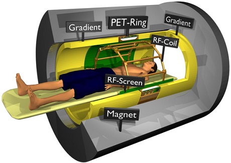
Fully integrated whole body PET/MRI system using a split gradient coil. Source: Philips
The researchers could identify in certain regions a mismatch between glucose metabolism related brain activation measured with PET and oxygenation related signals measured with MRI. Furthermore, information about functional connectivity in the brain could be derived from MRI and from dynamic PET data. These results help to further decipher the nature of brain function and are ultimately useful for basic research as well as clinical practice. The study “Simultaneous PET-MRI reveals brain function in activated and resting state on metabolic, hemodynamic and multiple temporal scales” by lead author Dr. Hans Wehrl of Prof. Pichler’s research team, is soon to be published in the journal Nature Medicine.
In PET imaging the distribution of a weakly radioactive substance is shown in cross-sections of the body, enabling doctors to see many different metabolic and physiological functions at work. Functional MRI (fMRI) allows researchers to depict changes in blood oxygenation that are associated with brain function. This measurement of functional active brain regions is also important for the planning of brain surgeries, where particular care must be taken in certain areas. The ability to collect different kinds of data from different scans simultaneously represents a major step forward in the fields using these technologies.
This research was conducted in close cooperation with the companies Siemens and Bruker and was supported from the German Research Foundation (DFG), the Wilhelm Schuler Foundation and the Werner Siemens Foundation.