Researchers at the School of Optometry at Indiana University (Bloomington, IN) have developed a new non-invasive and non-contact in vivo imaging system to test the human cornea with cellular-level resolution. This development offers tremendous opportunities to evaluate structural alterations that take place in different corneal diseases early in the process as well as monitor disease progression and evaluate treatment efficacy.
Being the most outermost element of the visual system puts the cornea at risk of infections, environmental damage, and a multitude of pathological conditions, including keratoconus, dry eye, and Fuchs endothelial corneal dystrophy. These inflictions can compromise the cornea’s integrity, leading to vision loss or even blindness. Advancements in in vivo confocal microscopy (IVCM) and optical coherence tomography (OCT) have greatly enhanced the ability to detect cellular characteristics of a multitude of corneal pathologies, but both methods have numerous drawbacks including low acquisition speed, patient discomfort, difficulty aligning the microscopic objective lens with the cornea, and the need for a highly trained professional operator.
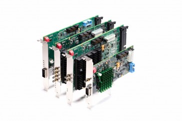
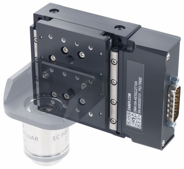
To overcome these limitations, the university researchers engineered a polarization-dependent optical coherence microscope (POCM) system. It enabled alignment with the human eye by integrating a bite bar, a headrest, and an in-line fixation target to stabilize the participant's head. Image signals were recorded with a customized spectrometer, equipped with a 250kHz line-scan camera set at 2048 pixel resolution connected to a BitFlow Axion-CL (AXN-PC2-CL-1xE) Camera Link 2.0 compliant frame grabber, enabling an imaging depth of ∼1 mm with a sensitivity greater than 97 dB. Highly deterministic, the Axion-CL supports one base, medium, full or 80-bit CL camera and clocks up to 85 MHz, along with offering the integration convenience of Power over Camera Link (PoCL). Synchronization between the scanners and the BitFlow frame grabber was achieved through a four-channel analog output data acquisition card.
Several strategies were applied to overcome motion artifacts inherent in other cornea imaging technologies. First, the camera's field of view was decreased from 1 × 1 mm to 0.5 × 0.5 mm, thus reducing the sampling points from 1000 × 1000 A-scans to 500 × 500 A-scans. This step yielded a four-time reduction in the acquisition time -- from 4 seconds to 1 second -- overcoming eye movements and motion artifacts. Custom programs in LabVIEW 2017 and MATLAB were also developed for data acquisition and processing. A flattening algorithm was applied to data sets to numerically remove the curvature of the cornea and correct for the axial motion artifacts, thus enabling the visualization of cellular features in enface images.
The proposed POCM enables non-invasive and non-contact in vivo imaging of all layers of the human cornea with a cellular-level resolution, which offers opportunities to evaluate structural alterations that take place in different corneal diseases early in the process. POCM has the potential to become a viable clinical tool for ophthalmologists, clinicians, and researchers to thoroughly examine the cornea in normal and pathological conditions.
Learn more at www.bitflow.com.