


Over the centuries, fossils have been collected and archived by museums, colleges and universities, and many private individuals all over the world. In some cases, however, a simple photograph may have been all that was available to others that might have wanted to view the specimens.
Often, such photographs rarely revealed many details about the fossils themselves, especially since no formal photographic methodology was adopted that might have enabled researchers to make valued judgments about them. Now, however, that’s about to change, thanks to Michael Eklund, Research Associate at the University of Texas at Austin, Vertebrate Paleontology Laboratory and ThinklabZ. Eklund has developed a technique called “Progressive Photonics” to record images of such fossils in a form that can greatly increase data yield and can be easily reproduced by researchers around the world.
Progressive Photonics
Progressive Photonics involves photographing paleontological specimens with the best of modern DSLR technology as well as greatly improved light sources to create a systematic and sequential series of images using different parts of the electromagnetic spectrum. It is a recording process designed to be easy to follow, relatively inexpensive, and one that produces far better specimen information. Interpretation of the specimens through new and better imaging techniques will benefit students, researchers, collection managers, collectors and the science as a whole.
ThinklabZ’ Mike Eklund says that his technique substantially improves data yield in three main problem areas that researchers currently face when examining fossil specimens.
First, many specimens are difficult to see because the color and/or texture of the specimen is too similar to the matrix surrounding it. Second, the visible spectrum does not enable researchers to observe the presence of many soft tissues, such as skin, scales, feathers, connective tissue or stomach contents that react better (by fluorescing) under non visible (UV or IR) light stimulation. Lastly, it may be that extensive repair work has been carried out on the specimen over the years. But with few records documenting such repairs, it may be challenging for researchers to determine the difference between biological evidence versus man made repairs or enhancements. Progressive Photonics, however, allows researchers to capture subtleties that typically go unnoticed.
True repeatability of results
“In the Progressive Photonics system, once the photographic composition, shot settings and focus are set, the specimens are not moved under the camera until the complete series of shots are completed. Only the light type, angle, filtering and descriptive tools change between images. A series of pictures are then captured so that it is easy to detect minute differences and details in the specimen characteristics. There is zero post processing or Photoshopping on any of the images because of the variability in the viewers’ perspective as well as variables in color calibration. This formula is what leads to true repeatability of results required for proper scientific documentation.” said Eklund.
The first image that is taken of each specimen is used as a setup shot from which later images of the specimen can be referred. As such, an image of the specimen is captured under direct lighting (direct overhead and shadowless) conditions with a label indicating the name and number of the specimen, as well as image scaling devices that enable researchers to judge both the size of the specimen, the angle of the lighting and the type of lighting that was used. To assist researchers in ensuring that the color of the digital images can be interpreted and reproduced accurately, a Munsell palette color calibration card is also present in the setup shot.
The second image captured of the specimen is taken using the same direct lighting but with most of the various labels and tools removed to provide a more uncluttered image.
Next, due to the fact that direct lighting alone may not reveal key features of the specimen, other images are then captured using multiple oblique and polarized lighting techniques. Typically, the images are taken with the light source illuminating the sample at four ninety degree angles. A special 1cm scale cube is placed on or alongside the specimen, enabling researchers to clearly understand the direction of the light source that was employed to illuminate the specimen when they review the images.


Images captured of a fossilized shark clearly demonstrate the effectiveness of the indirect lighting technique versus that of direct lighting.
“Images captured of a fossilized shark clearly demonstrate the effectiveness of the oblique lighting technique. While the direct lighting technique does provide a good indication of the outline of the specimen without any shadowing, the three dimensional detail and specific features such as its vertebral column are highlighted by the oblique technique, clearly delineating the sample from the background. Because it would be impossible to predict what features would best be highlighted by a specific orientation of lighting, the four images ensure that there is a strong possibility that one or more of the images will provide the optimum results,” said Eklund.
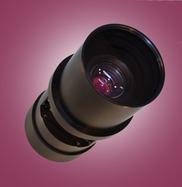
Visible light images are also captured of a specimen using an SLR camera fitted with a PC052 Circular polarizer from Midwest Optical Systems. The polarizer reduces the specular glare by passing only the light polarized in the direction perpendicular to the reflected light. In addition, it helps improve contrast, increase color saturation and help highlight details in a sample. In practice, an image of the sample is first captured with the polarizer disengaged, meaning with maximum glare. The polarizer is then rotated until the glare is reduced as much as possible and a second image of the sample is taken.
The role of UV imaging
Aside from visible light, the Progressive Photonics methodology also specifies that images of the specimen are captured in the ultraviolet radiation spectrum between 254 -400 nanometers. The UV imaging is used to capture the fluorescent reaction of the different minerals in specimens and surrounding matrix.

Unlike the images captured in the visible spectrum, when the specimen was illuminated with UVA light, the image captured by the SLR camera fitted with a UV filter clearly shows the gill arches and other specimen detail. In this shark image, an LA120 light balancing filter was used to reduce the violet dominance enabling features to be highlighted that may otherwise have been hard to interpret with the artificial skewed color.
The UV spectrum has long been understood to generate the broadest and most aggressive fluorescing reactions in elemental/mineral diagnostics. Depending upon the specimen itself, images will be captured in a number of UV ranges – notably the Ultraviolet A (or UVA) range from 320–400nm, the Ultraviolet B (or UVB) range from 280–320nm or the Ultraviolet C (or UVC) range from 100–280nm. To capture the visible light reaction of the specimen to UV excitation, the SLR camera is fitted with the appropriate UV filters from Midwest Optical Systems.
“The image of the shark is an outstanding example of the effectiveness of the UV imaging technique. Unlike the images captured in the visible wavelength, when the specimen was illuminated with UVA light, the image captured by the SLR camera fitted with a UV filter clearly shows new details of the gill arches, vertebral column and denticles. Due to the fact that the first UV image of shark specimen has an overwhelming dominance of violet, which may obfuscate some of the subtleties, an LA120 light balancing filter, also from Midwest Optical Systems, was used to reduce the violet tone, enabling features to be better highlighted that may otherwise have been concealed,” said Eklund.
The differences in specimens illuminated using different types of UV light can reveal some interesting facts about a sample. Images of an Asian rhino captured in visible light provide no indication of what might be the original specimen and what parts may have been modified in the past, perhaps to enhance the look of the piece.

Images of an Asian rhino captured in visible light provide no indication of what might be the original specimen and what parts may have been modified in the past.
 of artistic plaster fill and sculpture")
By illuminating the specimen with UVC light and applying a Midwest Optical Systems LA120 filter, however, the different minerals in the rhino skull become immediately obvious providing researchers with an indication that the skull was reconstructed and contains major portions (all dark areas) of artistic plaster fill and sculpture.
But by illuminating the specimen with UVC (short wave 254nm) light, the different materials in the rhino skull become immediately obvious, providing researchers with an indication that the skull may have been assembled from multiple disparate pieces and also have paint, plaster or adhesive present.
Known, specific and repeatable details
According to ThinkLabZ’ Eklund, what is important about the Progressive Photonics technique is that the images recorded by the rigorous methodology and complete documentation detail are known, specific and repeatable by anyone, and can be used and shared with total confidence by researchers anywhere in the world. He believes that the technique will provide researchers across the globe with a wealth of information that was previously unattainable simply through examining images of specimens taken under random lighting conditions.
Written by Dave Wilson, Senior Editor, Novus Light Technologies Today