


The molecular mechanism underlying chromosome condensation, one of the key steps in cell division, has been discovered from three-dimensional (3-D) refractive index (RI) reconstructions of individual mitotic cells. Writing in Nature Communications, the authors describe using the Tomocube HT-1S holotomographic microscope to quantify the structural and biochemical parameters of the cytoplasm and chromosomes within the individual mitotic cells.
The measurements, including the volume, concentration, and dry mass of the cell, as well as subcellular organelles, allowed the Korean research team to show that the asymmetric dimethylation at Arg2 on histone H3 (H3R2me2a) by protein arginine methyltransferase 6 (PRMT6) recruits the chromosomal passenger complex (CPC) to the chromosome arms and leads directly to chromosome condensation.
According to Aubrey Lambert, Tomocube’s Chief Marketing Officer, “Maintaining fidelity of mitosis is essential for life on Earth. However, despite being first described in the late 1870s, a complete description of the governing molecular mechanisms for the dramatic changes in chromosome organisation during chromosome condensation has remained elusive. The results of this latest study provide exquisite molecular insight into the mechanism by which the timely and temporal regulation of PRMT6 activity precisely confers flawless genetic inheritance on mitotic chromosomes.”
Chromosome condensation is one of the key steps during mitosis. The cell’s genetic material within the chromatin is compacted by a factor of two to three in mammalian cells to ensure that cell division is feasible within the cell space before the identical sister chromatids are separated from each other and paired along their entire length.
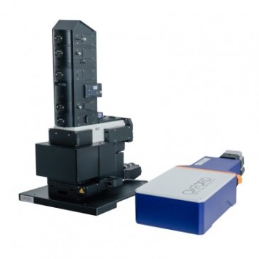
The Tomocube holotomography microscope delivers quantitative, nanoscale, real-time, 3-D images of individual living cells quickly and simply without any sample preparation. The holotomography images also deliver vital information on unique cell properties, including cell volume, shapes of sub-cellular organelles, cytoplasmic density, surface area, and deformability. Although not used in the chromosome condensation study, the latest HT-2 model combines the quantitative phase imaging (QPI) approach of label-free, 3-D refractive index (RI) tomography with 3-D fluorescence imaging. Winner of the Microscopy Today 2019 Innovation Award, this microscope enables long-term tracking of specific targets in live cells while minimising stress. The capability to easily deliver holotomography and fluorescence correlative analysis in 2D, 3D and 4D will enable researchers and clinicians to open new frontiers in bioscience and better understand, diagnose, and treat disease.
Reference
Seul Kim, Nam Hyun Kim, Ji Eun Park, Jee Won Hwang, Nayeon Myung, Ki-Tae Hwang, Young A Kim, Chang-Young Jang & Yong Kee Kim. PRMT6-mediated H3R2me2a guides Aurora B to chromosome arms for proper chromosome segregation. Nature Communications 11:612 (2020). https://doi.org/10.1038/