


Approximately 1.7 million people in the US receive traumatic brain injuries every year. The most common forms of traumatic brain injury are suffered by athletes, members of the military and those involved in motor vehicle collisions or occupational injuries. According to the US Centres for Disease Control and Prevention, these injuries cost the US health care system an estimated $60 billion USD per year.
According to a University of California San Francisco (UCSF) study, existing medical imaging technology could help predict long-term brain damage in patients with traumatic brain injury. (This article is a summary of the article "Imaging Technique Could Help Traumatic Brain Injury Patients" by Leland Kim, senior public information representative at UCSF.)
A form of imaging called magneto-encephalo-graphy (MEG) imaging was used to analyse the brain scans of traumatic injury patients. MEG imaging uses the magnetic fields produced by naturally-occurring electric currents in the brain to map the brain’s activity. The authors of the study discovered that long-term brain damage could continue years after even a mild brain injury. The senior author, Pratik Mukherjee, MD, PhD, associate professor in residence at the UCSF School of Medicine, said that they found a correlation between the MEG images and abnormal brain activity. The study was published on 19 April 2013 in the Journal of Neurosurgery.
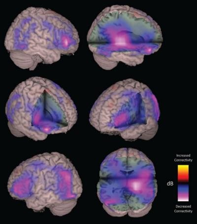
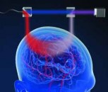
Magneto-encephalo-graphy (MEG) imaging shows brain scans of a patient with traumatic brain injury (TBI). Areas in pink and purple show reduced connectivity between parts of the patient’s brain.
MEG signals were measured for the first time in 1968. However, only recently has the technology been used widely for patients with traumatic brain injuries. A typical magnetic resonance imaging (MRI) scan uses magnetic field and radio wave energy to give a static image of the brain. MEG imaging provides richer information than an MRI. A MEG scan uses a computer algorithm to determine the areas of abnormality.
This study was supported by Dana Foundation Brain and Immuno-imaging Program, UCSF Research Evaluation and Allocation Program and US National Institutes of Health (Grant Nos. R01 NS060776 and RC2 NS069409). Co-authors include: Phiroz E. Tarapore, MD, and Geoffrey T. Manley, MD, PhD, of the UCSF Department of Neurological Surgery; Anne M. Findlay, BS, MA; Sara C. LaHue, BA; Hana Lee, MPH; Susanne M. Honma, RT; Danielle Mizuiri, BS; Tracy L. Luks, PhD; and Srikantan S. Nagarajan, PhD; all of the UCSF Department of Radiology and Biomedical Imaging.