


Light technologies are playing an increasingly significant role in diagnosing and treating many types of cancer. PET scans, CT scans, MRIs and endoscopy have been the traditional methods used in recent years, with rapid advances being made in research institutes around the world. Some of the recent advancements include 3D microscopy, combining light with an anti-malaria drug, using a nanoparticle agent for optical imaging, and a new molecular imaging technology.
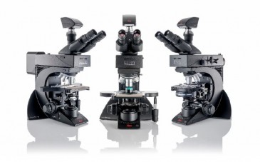
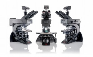
3D microscopy
In a study in Sweden, researchers used a 3D light sheet microscopy method to examine tumor tissue in three dimensions and found that it’s more accurate than 2D methods. The technique, as described in “3D Microscopy Gives More Accurate Cancer Diagnosis,” involves making tissue transparent and then reproducing it in three dimensions using the light-sheet microscope. The researchers studied stored samples from patients with bladder cancer, and were able to accurately diagnose tumors by recreating the 3D blood system feeding the tumors.
Fighting cancer with anti-malarial drug
The anti-malaria drug artemisinin is being tested by scientists from the National University of Singapore in combination with aminolaeyulinic acid, which is a photosensitizer. It’s been found to kill colorectal cancer cells and suppress tumor growth 10 times as much as if artemisinin were administered alone. This therapy could also have fewer side effects.
The study, which is described in “Light Combined with Anti-Malaria Drug Shows Promise for Treating Cancer,” showed that artemisinin is activated by heme, an iron-containing compound of the porphyrin class that forms the nonprotein part of hemoglobin and some other biological molecules. When parasites digest hemoglobin, they release heme, which activates artemisinin in the parasites, thus attacking multiple proteins that they need to survive. The researchers found that cancer cells have higher heme levels than non-cancer cells. So when heme is activated in cancer cells, artemisinin attacks more than 300 proteins, many of which are important for the survival of cancer cells.
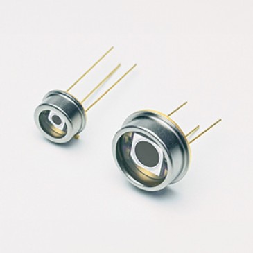
MRI gets a new agent
Magnetic resonance imaging has long been used to successfully diagnose cancer cells, and yet researchers in India have taken MRIs to the next level. They developed a less-toxic, nanoparticle-based contrast agent that produce particularly bright images, which means that cancer cells can be identified with greater accuracy. Because a minimal amount of contrast agent is needed, and the researchers found that toxicity could be lowered even more by coating the nanoparticles with silica.
In the article “Nanoparticle Agent for MRI and Optical Imaging of Cancer Cells,” the lead researcher indicates that they are optimizing the size and morphology of the particles by varying parameters, so contrast agents will be able to be developed with low toxicity, higher reflexivity and longer lifetime, and will be optimized for clinical use.
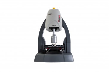
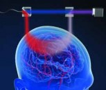
Photoacoustic microscopy mixes light and sound
One of the great challenges in breast-conserving cancer surgery (lumpectomies) is to not remove any more of the breast tissue than is necessary. Sometimes too much is left behind, however, making a second surgery necessary.
Researchers at Caltech used photoacoustic microscopy, exciting a tissue sample with a low-energy laser, which causes the tissue to vibrate. As described in “Light and Sound Microscopy Technique Reduces Cancer Surgeries,” the system measures the ultrasonic waves emitted by the vibrating tissues. Cancerous tissue tends to have larger nuclei and more densely packed cells, and this microscopy method can determine the size of the nuclei and the packing density of the cells. One of the benefits of this method is speed. A sample can be analyzed in about three hours compared to the seven it takes with traditional microscopy. And with a laser with faster pulse repetition and parallel imaging, the time could be cut down to 10 minutes or less, according to the lead researcher, making this technology appropriate for clinical settings.
Written by Anne Fischer, Managing Editor, Novus Light Technologies Today